Melanoma is the most serious type of skin cancer: aggressive forms of the disease can grow and spread rapidly to other parts of the body, highlighting the urgent need for new treatments.
But over the past few years, genetic analysis has revealed that melanoma cells –
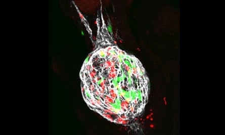
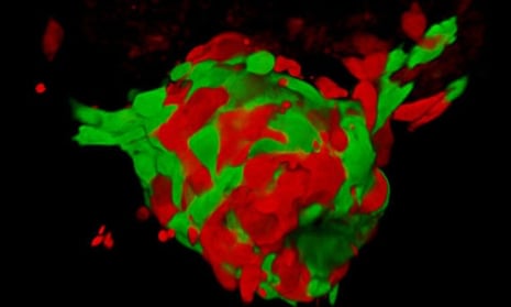
like many other tumour cell types – are not all created equal. In fact, the cells that make up a melanoma come in different genetic ‘flavours’ – two of which are shown in red and green in the image above. Until today, these were thought to have fairly regimented roles in the growth and spread of the disease.
But a team of Cancer Research UK-funded scientists from the University of Manchester – led by Dr Claudia Wellbrock – has found that these genetically distinct groups of cells may actually work together co-operatively, to break away from the tumour and spread around the body.
And their latest findings – published in the journal Cell Reports – have emerged following work with tiny transparent zebrafish (see video) to find out more.
Studies have shown that melanoma cells produce different amounts of a protein, called MITF, and behave in different ways. MITF is a protein known as a transcription factor, responsible for switching a cell’s genes on or off. Tumour cells packing a lot of MITF – coloured red in images here – tend to grow more quickly, but are less able to spread.
On the other hand, cells carrying low amounts of MITF – like those in green – are much more mobile.
But tumours aren’t made up of just one type of cell. To find out what happens in a tumour, the Manchester team mixed the cells together and watched how they behaved.
When these cells formed a tumour in zebrafish, they saw the green cells physically helping the slower-spreading red cells break away from the tumour and spread.
When they looked more closely, the team found that the green cells often lead the way, encouraging red cells to move as part of an invasive single file of cells.
As with many solid tumours, in order to spread, melanoma cells have to be able to navigate the complex meshwork of molecules and protein fibres that surround the tumour.
To do this, they often deploy an arsenal of enzymes that can melt away this meshwork. These enzymes are known as ‘matrix metalloproteinases’, or MMPs for short.
When the Manchester team looked at mixtures of the red and green tumour cells in zebrafish, they also relied on MMPs to spread, and the researchers believe that the green cells may be using MMPs to clear a path for the red cells to follow.

These mixtures of cells were also better at laying down important protein ‘tracks’ – shown in white, above – that surround the red and green tumours.
The researchers found that the red cells relied on these tracks to escape and move away from the tumour. And when the team engineered the green tumour cells to stop making one of the key components of these tracks – a protein called fibronectin – the red cells were no longer able to move away from the tumour.
This latest study provides important clues about how melanoma cells help each other spread around the body. And it reinforces how tumours are made up of a complex mixture of cells and molecules.
By looking at this mixture, and analysing how tumour cells interact with each other, scientists are piecing together the crucial events that help melanoma cells spread.
And by doing this, we move a step closer to understanding how to stop it.
Dr Nick Peel is a science media officer at Cancer Research UK


Comments (…)
Sign in or create your Guardian account to join the discussion