

Summary: Researchers have identified specific brain networks that helps us associate objects with their names. The study sheds light on how the brain connects meaning to words and could help explain why people with neurodegenerative diseases often have problems naming every day objects.
Source: UT Houston.
Scientists at The University of Texas Health Science Center at Houston (UTHealth) have identified the brain networks that allow you to think of an object name and then verbalize that thought. The study appeared in the July issue of BRAIN. It represents a significant advance in the understanding of how the brain connects meaning to words and will help the planning of brain surgeries.
Their discovery could help explain why people with neurodegenerative disease often forget the names of objects. An estimated 5.7 million Americans of all ages have Alzheimer’s dementia. Described as the tip-of-the-tongue phenomenon in healthy individuals, the inability to recall the name of items is a condition called anomia.
“Object naming has been a core method of study of anomia, but the processes that occur when we come up with these names, generally in less than a second, are not well understood. We mapped the brain regions responsible for naming objects with millimeter precision and studied their behavior at the millisecond scale,” said Nitin Tandon, M.D., the study’s senior author and a professor in the Vivian L. Smith Department of Neurosurgery at McGovern Medical School at UTHealth.
“The role of the basal temporal lobe in semantic processes has been underappreciated. Surgeons could use this information to design better approaches for epilepsy and tumor surgery, and to reduce the cognitive side effects of these surgical procedures,” said Tandon, who is also the director of the epilepsy program with the Memorial Hermann Mischer Neuroscience Institute-Texas Medical Center and a member of the faculty at The University of Texas MD Anderson Cancer Center UTHealth Graduate School of Biomedical Sciences.
Tandon added that this study is of particular value as it produced convergent maps with three powerful techniques: electrophysiology, imaging and brain stimulation.
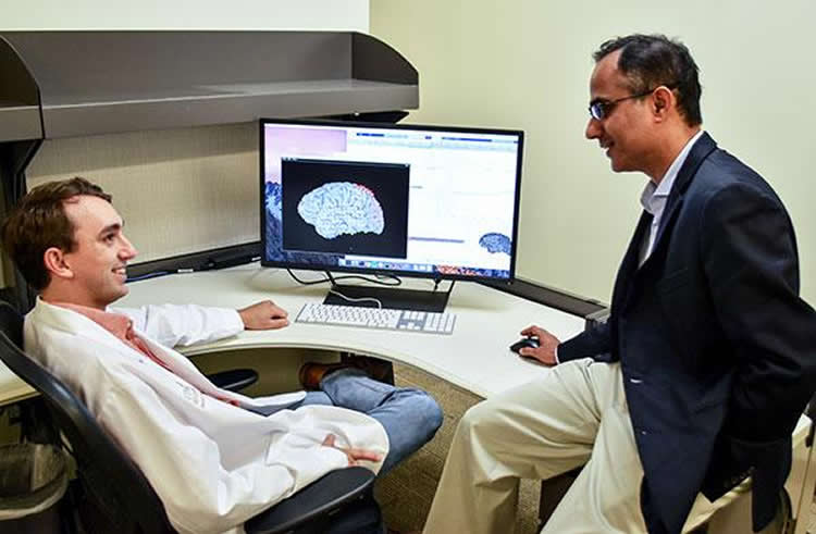
While their brain activity was being monitored for epileptic seizures, 71 patients were asked to look at a picture of an object and identify it and/or asked to listen to a verbal description of an object and name it. Much like explorers mapped the wilderness, the researchers used these brain data to map out the brain networks responsible for certain processes.
With the aid of both electrocorticography and functional magnetic resonance imaging, researchers zeroed in on the specific brain regions and networks involved in the naming process. This was then confirmed with a pre-surgical mapping technique called direct cortical stimulation that temporarily shuts down small regions of the brain.
“The power of this study lies in the large number of patients who performed name production via two different routes and were studied by three distinct modalities,” said Kiefer Forseth, the study’s lead author and an M.D./Ph.D. student at MD Anderson UTHealth Graduate School.
Tandon and Forseth’s coauthors include Cihan Mehmet Kadipasaoglu, M.D., Ph.D., and Christopher Richard Conner, M.D., Ph.D., who both conducted their dissertation research in the Tandon lab at MD Anderson UTHealth Graduate School.
Funding: Funding provided by NIH/National Institute on Deafness and Other Communication Disorders, National Center for Research Resources Clinical and Translational Science Award.
Source: Rob Cahill – UT Houston
Publisher: Organized by NeuroscienceNews.com.
Image Source: NeuroscienceNews.com image credited to Rob Cahill, UTHealth.
Original Research: Abstract for “A lexical semantic hub for heteromodal naming in middle fusiform gyrus” by Kiefer James Forseth, Cihan Mehmet Kadipasaoglu, Christopher Richard Conner, Gregory Hickok, Robert Thomas Knight, and Nitin Tandon in Brain Published July 1 2018.
doi:10.1093/brain/awy120
[cbtabs][cbtab title=”MLA”]UT Houston”Brain Networks Responsible for Naming Objects Identified.” NeuroscienceNews. NeuroscienceNews, 15 August 2018.
<https://neurosciencenews.com/object-naming-networks-9704/>.[/cbtab][cbtab title=”APA”]UT Houston(2018, August 15). Brain Networks Responsible for Naming Objects Identified. NeuroscienceNews. Retrieved August 15, 2018 from https://neurosciencenews.com/object-naming-networks-9704/[/cbtab][cbtab title=”Chicago”]UT Houston”Brain Networks Responsible for Naming Objects Identified.” https://neurosciencenews.com/object-naming-networks-9704/ (accessed August 15, 2018).[/cbtab][/cbtabs]
Abstract
A lexical semantic hub for heteromodal naming in middle fusiform gyrus
Semantic memory underpins our understanding of objects, people, places, and ideas. Anomia, a disruption of semantic memory access, is the most common residual language disturbance and is seen in dementia and following injury to temporal cortex. While such anomia has been well characterized by lesion symptom mapping studies, its pathophysiology is not well understood. We hypothesize that inputs to the semantic memory system engage a specific heteromodal network hub that integrates lexical retrieval with the appropriate semantic content. Such a network hub has been proposed by others, but has thus far eluded precise spatiotemporal delineation. This limitation in our understanding of semantic memory has impeded progress in the treatment of anomia. We evaluated the cortical structure and dynamics of the lexical semantic network in driving speech production in a large cohort of patients with epilepsy using electrocorticography (n = 64), functional MRI (n = 36), and direct cortical stimulation (n = 30) during two generative language processes that rely on semantic knowledge: visual picture naming and auditory naming to definition. Each task also featured a non-semantic control condition: scrambled pictures and reversed speech, respectively. These large-scale data of the left, language-dominant hemisphere uniquely enable convergent, high-resolution analyses of neural mechanisms characterized by rapid, transient dynamics with strong interactions between distributed cortical substrates. We observed three stages of activity during both visual picture naming and auditory naming to definition that were serially organized: sensory processing, lexical semantic processing, and articulation. Critically, the second stage was absent in both the visual and auditory control conditions. Group activity maps from both electrocorticography and functional MRI identified heteromodal responses in middle fusiform gyrus, intraparietal sulcus, and inferior frontal gyrus; furthermore, the spectrotemporal profiles of these three regions revealed coincident activity preceding articulation. Only in the middle fusiform gyrus did direct cortical stimulation disrupt both naming tasks while still preserving the ability to repeat sentences. These convergent data strongly support a model in which a distinct neuroanatomical substrate in middle fusiform gyrus provides access to object semantic information. This under-appreciated locus of semantic processing is at risk in resections for temporal lobe epilepsy as well as in trauma and strokes that affect the inferior temporal cortex—it may explain the range of anomic states seen in these conditions. Further characterization of brain network behaviour engaging this region in both healthy and diseased states will expand our understanding of semantic memory and further development of therapies directed at anomia.


