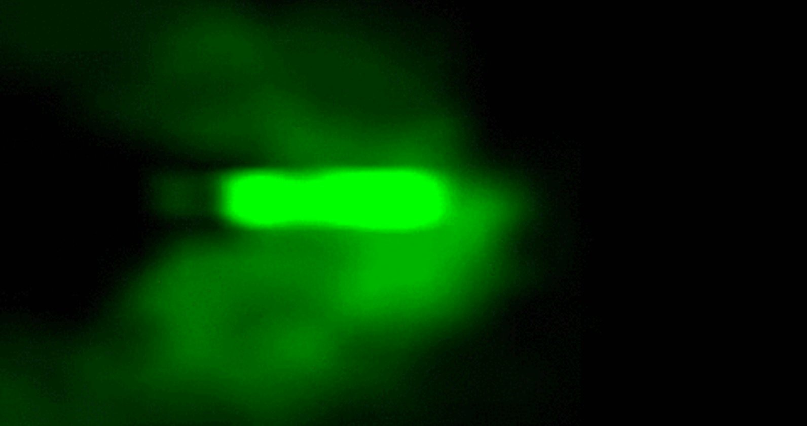
Scientists Capture Time-Lapse of an Immune Cell Moving Through the Body
In April of 2018, a team of scientists made an imaging breakthrough that allowed them to capture this incredible video of an immune cell moving through the tissues of a zebra fish’s inner ear, picking up sugar particles as it goes along.
The video resurfaced today when it exploded in popularity on the r/videos subreddit, where it’s received some 10k upvotes over the past ten hours. And not without reason: the fact that this kind of footage exists is genuinely mind-blowing.
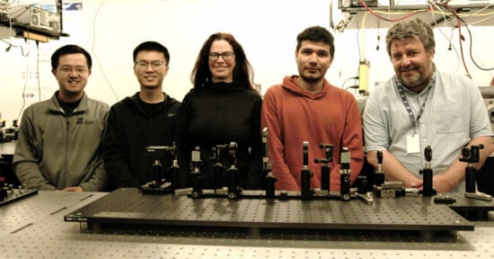
This breakthrough in microscopic imaging was made by a team of researchers led by Physicist Tsung-Li Liu, who combined two different imaging techniques into a hybrid that allows them to “study a variety of delicate subcellular events in vivo.” In other words: they can capture highly detailed 3-D images of living cells moving through living tissue.
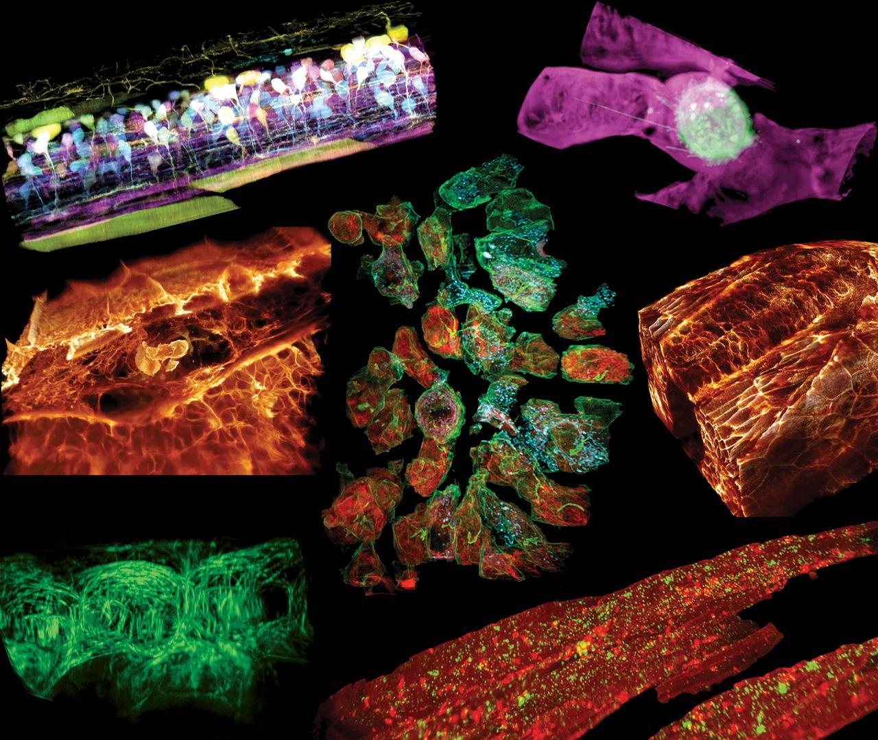
The example above shows an immune cell buzzing around a zebrafish inner ear, but they were able to capture many more examples of subcellular activity, like neural circuits developing in a zebrafish spinal chord and a cancer cell migrating through a blood vessel:
The technique they developed is described in technical detail in the journal Science, but in summary, the researchers developed a new kind of microscope by combining lattice light-sheet microscopy (LLSM) with adaptive optics (AO) to create a system that can capture 3D ‘movies’ of specific structures in vivo at an unprecedented level of detail.
The lattice light-sheet microscope “rapidly and repeatedly sweeps an ultrathin sheet of light” through the subject—collecting imaging data and building a 3D movie—while the adaptive optics system “measures sample-induced distortions […] and compensates for these by changing the shape of a mirror to create an equal but opposite distortion.”
That allows the scientists to go from an image that is, a researcher Eric Betzig told HHMI, “too damn fuzzy” to a high-resolution 3D time-lapse that captures the activity as it’s happening. The immune cell video, for example, is made up of 90-minutes worth of images taken every 13 seconds.
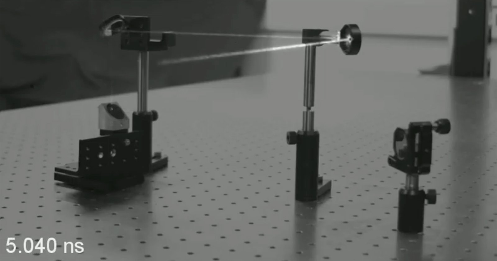
This first iteration of the microscope was, unfortunately, massive. According to HHMI, it takes up about a 10-foot-long table. But it was a proof of concept that researchers were already working on shrinking down back in 2018.
As this technique and technology advances (and shrinks) we’ll begin to see many more amazing videos like the ones above: bona fide peeks into the inner workings of living cells.