Search this site
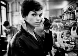
Photography Job You’ve Never Heard Of: Medical Photographer
This article is part of our series on unusual and inspiring photography jobs. Today, we’re highlighting medical photographer Eneil Simpson.

“You get the picture the first time, or you don’t get it at all,” the ophthalmology and medical photographer Eneil Simpson says of working in the operating room. “You can’t stop the procedure for a better picture, and there are no do-overs.”
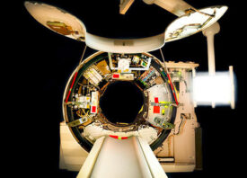
To get the shot he needs, he’s looked over shoulders, when permitted, and ducked under arms, all while remaining invisible. Most of the time, he uses a compact and portable two-step ladder positioned by the patient’s feet. He’s stood in the same place for a dozen hours at a time, documenting brain and heart cases, to name a few, from start to finish.

Simpson got his start as a medical photographer while in between jobs, after his ophthalmologist suggested he give it a try. To this day, the eye remains his primary area of expertise; “Most of my work is in the ophthalmology clinic,” he explains. “I only go into the OR on my time off, after having requested permission to photograph a case that might be interesting.”

From those early days in the field, Simpson felt at home in medical settings, even in cramped spaces where getting a photo–and staying out of everyone’s way–proved to be difficult. “I felt comfortable in the OR from the beginning,” he remembers. “I wear scrubs like the doctors do. My conduct in the OR is to be professional at all times. Touch nothing unless asked.


“I like the surgeons to not be aware that I am even in the room. In the OR, I am a guest. I try to find a spot that will not interfere with the movement of the techs in the room. I pay attention to what is going on around me and am prepared to move if need be.”
Once he’s captured the images in the OR–sometimes hundreds in one go–Simpson gives them to the surgeon, and they’re ultimately used in lectures for educational purposes. As part of his day job in the eye clinic, his photos end up in the patients’ records and are used when residents present cases in grand rounds.

The ophthalmology work is also different from the surgical cases in the sense that Simpson gets to meet the patients. “I have gotten to know patients as they come for their visits at the eye clinic, and the eye patients see their photos as we take them,” the photographer tells us.
He stresses that his job is never to treat or diagnose patients, but the career path has led him in unexpected directions as his knowledge of the field has grown; among other milestones, Simpson co-authored a medical paper, along with one of his attending doctors. He’s also been contacted by graduate students looking to enter the field of medical photography.

Medical photography is a niche genre, and in some cases, Simpson says surgeons might opt to take pictures themselves using a cell phone. Still, despite all the long hours and unpredictable work environment–and perhaps because of them–Simpson’s thankful his life took the unexpected turn into medical photography.
“What can I say, seeing inside of the human body is truly fascinating,” he tells us. “I have photographed the eyes of newborn babies and watched as they grew and now are entering college. My oldest patient is 103 years old.”

All images by Eneil Simpson